 |
Macro method (wintrobe method) for estimation of packed cell
volume (pcv) or hematocrit
|
Macro method (wintrobe method) for estimation of packed cell volume (pcv) or hematocrit
PACKED CELL VOLUME (PCV):-
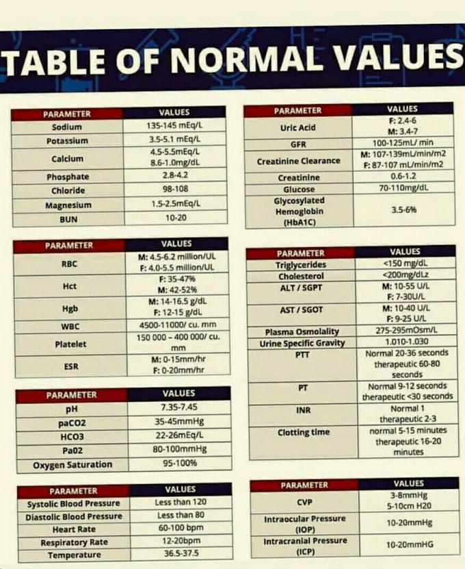
The PCV or Hematocrit is a percentage of the total volume of whole blood
occupied by packed red blood cells, when a known volume of whole blood is
centrifuged at a constant speed for a constant period of time. Value thus
obtained is used in the determination of the red cell indices: mean corpuscular hemoglobin (MCH), mean corpuscular hemoglobin concentration (MCHC) and the
mean corpuscular volume (MCV).
Macro method (wintrobe method):-
 |
Macro method (wintrobe method) for estimation of packed cell
volume (pcv) or hematocrit
|
Apparatus : Wintrobe's Tube
Specimen
(Plain capillary tubes may be coated with heparin by filling them with 1:1000 dilution of heparin and drying at 56°C).
Fill the capillary tube two-thirds full either with well
mixed venous blood or directly from a capillary puncture. Seal one end of the
capillary tube with modelling clay (plasticine).
The filled tubes are then placed in the microhaematocrit centrifuge and spun at 12,000 g for 5 minutes. Place the spun tube into a specially designed scale, and read the PCV as a percentage. Alternatively, PCV can be calculated as for the
ii. Buffy coat can be prepared for other tests.
iii. By seeing the colour of plasma we can know about
some of the pathological conditions e.g. in jaundice
it is yellow, in haemolysis it is pink, in hyperlipidaemia
it is milky.
The filled tubes are then placed in the microhaematocrit centrifuge and spun at 12,000 g for 5 minutes. Place the spun tube into a specially designed scale, and read the PCV as a percentage. Alternatively, PCV can be calculated as for the
Procedure
 |
| Heamorcrit by wintrobe's tube method |
- Fill the Wintrobe tube upto mark 10 with well mixed
anticoagulated blood (EDTA) by Pasteur’s pipette
free of air bubbles.
- Centrifuge the tube at 2000-2300 g for 30 minutes.
- After centrifugation layers are noted in the wintrobe tube as under (Fig. 50.2):
- Uppermost layer of plasma.
- Thin white layer of platelets.
- Greyish-pink layer of leucocytes.
- Lowermost is the layer of RBCs.
- Grey-white layer of leucocytes and plateletsinterposed between plasma above and packedRBCs below is called buffy coat.
OR
- With a long stemmed pasteur pipette fill the hematocrit tube up to the top mark, i.e., 100 mm, avoiding air bubbles. Centrifuge at 3000 r.p.m. for 30 minutes.
- The bottom of the tube thus contains packed red cells and on top it has a small buffy layer, consisting of the white blood cells and the platelets and a column of plasma above it.
- Without shaking the tube read the volume of packed red cells on the tube, expressed as a percentage of the whole blood.
- If the height of the blood column is not exactly 100 mm, a simple calculation can be made to correct this:
Note
the lowermost height of column of packed RBC layer and express it as percentage.
Packed RBC column height PCV % = Total blood column height x 100
Advantages of Macro Method
i. PCV and ESR can be measured simultaneously.ii. Buffy coat can be prepared for other tests.
iii. By seeing the colour of plasma we can know about
some of the pathological conditions e.g. in jaundice
it is yellow, in haemolysis it is pink, in hyperlipidaemia
it is milky.


If you have any queries related medical laboratory science & you are looking for any topic which you have have not found here.. you can comment below... and feedback us if you like over work & Theory
.
Thanks for coming here..