Liver Function
SECRETION OF BILE
FUNCTIONS OF THE LIVER
METABOLISM OF BILIRUBINJaundice (icterus)
1. Prehepatic2. Hepatic
3. Post-hepatic
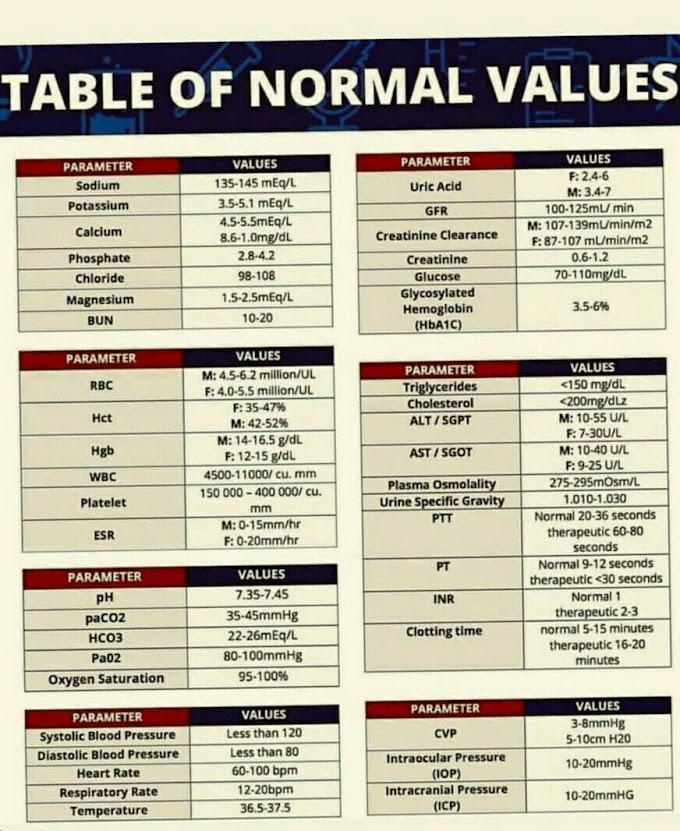
LIVER FUNCTION TESTS
Principle Reagents Method
Calculation
Example
Normal range Clinical Significance
Laboratory diagnose
Liver Function Tests
Calculation
Example
Normal range Clinical Significance
Laboratory diagnose
The liver is the largest organ in the body. It weigh about 1.5 kg in an adult. It is also the most active
and most complex organ. It lies in the upper right side of the abdomen beneath
the diaphragm. It has four lobes, the right one being the largest while the
left one is smaller and wedge shaped The other two lobes are very small and can
only be seen on the under surface of the liver.
The portal vein which enters the liver through the portal
tissue carries blood from the stomach, spleen, pancreas and intestines. The
hepatic artery carries the arterial blood to the liver. The hepatic vein leaves
the liver to carry blood to the inferior vena cava. The right and left hepatic
ducts carry bile to the gall bladder.
The liver is divided into many small circular units-lobules.
Each lobule is made of rows of cells—the hepatocytes, radiating from the
central vein.
SECRETION OF BILE
Bile is secreted by the liver cells, stored and concentrated
in the gall bladder, and then released into the duodenum where it helps to
emulsify dietary fat. Between meals the sphincter of Oddi in the gall bladder
is closed and it is opened at the sight, taste or smell of food.
When the gastric contents enter the duodenum, a hormone known as cholescystokinin and which is secreted by the intestinal mucosa, causes the gall bladder to contract, thereby making bile enter the duodenum.
Here the bile is essential for adequate reabsorption of fats and of vitamin K. Reduction of bile by bacteria produces bile salts (usually sodium or potassium) such as deoxycholic acid and lithocholic acids conjugated with glycine and taurine.
Bile is golden yellow or greenish coloured fluid that
contains cholesterol, bilirubin and bile salts. Much of the bile secreted into
the intestine is reabsorbed into the hepatic portal circulation, recycled in
the liver and re-excreted in the duodenum. Only small amount of bile is
normally excreted in the faeces.
FUNCTIONS OF THE LIVER
1. Excretion of bile The liver produces and excretes bile
into the intestine
2. Carbohydrate metabolism The main monosacharrides obtained
during digestive processes are converted into glycogen and stored in the liver.
When required, the glycogen is re-converted to glucose (Glycogenolysis).
Thus blood glucose level is maintained. But if the liver has used all the stored glycogen and blood glucose is below normal, the liver can convert protein and fat into glucose (gluconeogenesis).
Thus blood glucose level is maintained. But if the liver has used all the stored glycogen and blood glucose is below normal, the liver can convert protein and fat into glucose (gluconeogenesis).
3. Protein production Plasma proteins, namely, fibrinogen,
albumin and globulin (except gamma globulin) are manufactured in the liver. The
liver also produces transport proteins such as transferrin which binds and
transports iron, and haptoglobin which combines with free haemoglobin.
4. Synthesis of blood clotting factors Many of blood
coagulation factors, fibrinogen, prothrombin, factors V, VII, IX, X, XI and XII
are synthesised in the liver.
5. Storage In addition to glycogen and many vitamins, the
liver is the main organ of storage for iron.
6. Detoxication The ammonia released from amino-acid
deamination is detoxciated by being converted to urea for excretion by the
kidney. The liver also detoxicates drugs, metabolic products, hormones and
alcohols by oxidation, conjugation, methylation or reduction.
7. Metabolism of lipid The synthesis of cholesterol,
phospholipids, endogenous triglycerides and lipoprotein occurs mainly in the
liver, including the esterification of cholesterol.
8. Phagocytic activity
Worn out red blood cells as well as micro-organisms are removed by the Kupffer
cells which form a part of mononuclear phagocytic defence system (a part of
reticuloendothelial system, (RES)) of the body.
• METABOLISM OF BILIRUBIN
Bilirubin is mainly formed from the breakdown of haemoglobin
in the cells of the liver, spleen and bone marrow. The haem (iron porphyrin ) of the haemoglobin
molecule is firstly removed from the globin.
The porphyrin portion is then converted to biliverdin which is reduced to bilirubin. The bilirubin is insoluble in water, it is carried in the blood attached to albumin and it cannot be excreted by the kidney. This bilirubin is referred to as unconjugated (indirect) bilirubin.
The enzyme glucuronosyl transferase in the liver cells joins
(conjugates) glucuronic acid to bilirubin to form bilirubin glucuronides. This
bilirubin is called conjugated (direct) bilirubin and it is water-soluble and
non-toxic.
Conjugated bilirubin is passed from the liver into the gall
bladder through the bile duct, and into the intestine. Here the bilirubin is
deconjugated and reduced by bacterial enzymes to a colourless compound,
stercobilinogen (faecal urobilinogen).
This is oxidised to a brown pigmen, stercobilin, the main pigment of faeces Some of the stercobilinogen is re-absorbed via the portal vein back to the liver from where it re-enters the intestine in the bile and is excreted in the faeces. A small amount of the reabsorbed urobilinogen is carried in the blood through the liver and transported to the kidneys where it is excreted in the urine.
This is oxidised to a brown pigmen, stercobilin, the main pigment of faeces Some of the stercobilinogen is re-absorbed via the portal vein back to the liver from where it re-enters the intestine in the bile and is excreted in the faeces. A small amount of the reabsorbed urobilinogen is carried in the blood through the liver and transported to the kidneys where it is excreted in the urine.
Bile salt Together with the bile, bile salts enter the
duodenum and are found in small amounts in faeces and very little in urine. The
bile salts are reabsorbed via the portal vein, removed by the liver and
excreted in the bile. In obstructive jaundice, bile salts are present along
with bile pigments in the urine.
Jaundice (icterus)
This is the condition during which the patient has a yellow colouration of the eyes and some parts of the skin. The yellow pig. mentation is due to a raised level of bilirubin which is normally present in small amounts in the plasma. Jaundice may be classified into three types.
1. Prehepatic Due to
an excessive breakdown of red blood cells as in haemolytic anaemia, the
bilirubin load becomes too much for the liver to conjugate. The bilirubin is
therefore mostly unconjugated or indirect. summarises the metabolism
of bilirubin.
2. Hepatic As a result of diminished function of the liver
cells (due to damage) as in infective hepatitis, conjugation is inhibited; so
initially the bilirubin is indirect. As the cell damage affects the structure
of the liver, conjugated bilirubin becomes increased. Thus both types of
bilirubin are present in the sarum.
3. Post-hepatic When bilirubin which has been conjugated
cannot be excreted due to obstruction in the bile duct by either gall stone
within the lumen or by a tumour exerting pressure from outside, the bilirubin
"spills over" into the blood. This bilirubin is water-soluble
(conjugated) and can be excreted by the kidney. Table 5.1 shows the type of
bilirubin present in various conditions.
LIVER FUNCTION TESTS
These are routine tests performed in the investigation of a
liver disease. They include serum or plasma and urine bilirubin, urine
urobilinogen, plasma alkaline phosphatase, plasma alanine
Mix well and read absorbance at 660 nm (red filter)
Calculation
Note
1. One Caraway unit is the amount of amylase per 100 ml of serum/ plasma which will hydrolyse 5 mg starch in 15 mintues at 37°C. In the test, 2.5 ml of buffered substrate is used which contains 1 mg starch (400 mg/L). Incubation period is for 7.5 minutes. The amount of serum used is 0.1 ml. Therefore, the factor 400 is derived as follows:
2. Amylase International Units/Litre
(U/L) = Caraway units x 1.85
3. Avoid contamination of the test with saliva or sweat as
they contain amylase.
4. If serum amylase is more than 400 Caraway units, dilute the serum 1:5 and multiply the result
by the dilution factor.
Normal values
60-160 Caraway units
100-340 International units
Amylase in Urine Urinary amylase (aka diastase) is raised in
acute pancreatitis and may remain so even after the se rum level has returned to normal.
The method of measurement given here was originally described by Wohlgemuth in 1908, and modified by Harrison. It is a good example of urinary enzyme determination that provides a useful practical exercise for students.
The method of measurement given here was originally described by Wohlgemuth in 1908, and modified by Harrison. It is a good example of urinary enzyme determination that provides a useful practical exercise for students.
Principle
The amylase in urine breaks down starch into smaller compounds and finally to maltose. Starch gives blue colour with iodine but when it has been broken down to around 10 glucose units, it gives red colour, and at less than 5 glucose units, no colour is produced.
The amylase in urine breaks down starch into smaller compounds and finally to maltose. Starch gives blue colour with iodine but when it has been broken down to around 10 glucose units, it gives red colour, and at less than 5 glucose units, no colour is produced.
Specimen Collect a 24 hour urine specimen with thymol as
preservative.
Reagents
(a) Starch solution
(i) Stock solution Make a paste with 2 g of
soluble starch and small volume of distilled water; then wash
into 60 ml of boiling water. Add 10 g of sodium chloride, dissolve, cool and transfer
to a 100 ml volumetric flask and make up to mark with distilled water.
(ii) Working starch solution Dilute stock solution 1:20 with
distilled water.
(b) Iodine solution
(i) Stock solution
(approx. 0.05 M) Weigh 6.4 g of iodine into a 500 ml flask and add 10-12 g KI
(Analar, free from KIO) and 20 ml of distilled water. Stopper the flask, mix thoroughly to dissolve. Make to the mark with distilled water.
(ii) Working solution of iodine (approx.0.01M) Dilute stock
solution 1:5 with distilled water.
Method
1. Adjust pH of urine to less than 7.0 using concentration
2. Arrange a series of tubes as follows:
3. Incubate at 37°C for 30 minutes.
4. Cool immediately in cold water.
5. Add working iodine solution drop by drop from a pipette to
each tube, watching carefully for the appearance of a bluecolour.
6. Note the smallest
amount of urine whichdoes not give a blue colour.
Calculation Diastase index = ml of 0.1% starch digested by
1ml of urine at 37°C in 30 minutes
Example If on completion of test, a blue colour was first
noticed in tube 7, so the smallest volume of urine not showing a blue colour is
1.0 ml of 1:10 dilution in tube 6 (i.e., 0.1 ml) Therefore
Diastase index =
The diastase index should be multiplied by the total volume
of a 24 hour sample of mine.
Normal range Up to 3000 Caraway units for 24 hours
Note
Urinary amylase can also be estimated in the same way as
serum amylase on a 24 hour specimen.
Clinical Significance
Increased serum amylase (>1800 U/L) is diagnostic of acute
pancreatitis. The rise in serum amylase level is often brief and the level may
return to normal within 48-72 hours. Urinary amylase, on the other hand may
persist for a longer period.
Serum amylase is moderately increased (7401500 U/L) in
conditions such as renal failure, diabetic ketoacidosis, hypothermia,
perforated peptic ulcer or appendicitis.
Normal values may be obtained in chronic pancreatitis.Fibrocystic Disease of the Pancreas (Cystic fibrosis)
Normal values may be obtained in chronic pancreatitis.Fibrocystic Disease of the Pancreas (Cystic fibrosis)
This is a familial Mendelian recessive disease characterised
by abnormally high secretion of sodium and chloride by the various exocrine
glands of the body such as pancreas, salivary, peritracheal, sweat and lacrimal
glands as well as the glands of the small intestine and bile ducts.
Laboratory diagnosis
The principle is to demonstrate increased sodium and chloride in the sweat of patients. Screening test for sweat chloride depends upon hand imprints on agar or paper containing silver nitrate.Chloride precipitates with silver and intensity of the print is roughly proportional to the chloride precipitation. Chemical estimation is more reliable. Analysis of electrolytes in sweat is a most valuable investigation. Collection of sweat is carried out using:
(1) Methacholine chloride Injecting 2 mg of methacholine
chloride into the forearm and covering the area with filter paper. This
produces local stimulation of sweat. About 100-300 mg of sweat can be
collected. This method is now obsolete.
(2) Pilocarpine iontophoresis A current of 1.5 mA is passed
between the two electrodes for about 30 minutes. The positive electrode
contains 0.5% aqueous pilocarpine nitrate solution while the negative electrode
contains 1% aqueous sodium nitrate solution.
Ashless filter paper saturated with pilocarpine nitrate solution is placed under the surface of the positive electrode and under the surface of the negative electrode is a gauze saturated with sodium nitrate solution. The positive electrode is then strapped to the flexor surface and the negative electrode on the exterior surface of the forearm.
The area of the positive is washed with distilled water. Whatman No. 40 filter paper of a known weight is carefully placed over the area and covered with parafilm. The sweat is collected for about 30 minutes.
The paper is then
removed, weighed, placed in a flask. The electrolytes are eluted with 10 ml
distilled water. The concentration of sodium and chloride ions is measured in
the usual way A machine called the cystic fibrosis analyser, has been developed
to stimulate sweat by a pilocarpine iontophoresis and to measure the sodium and
chloride concentrations by conductivity meter.
(3) Wescar sweat collection system This system is also based
on pilocarpine iontophoresis but uses a unique collection device-a heated cup
which reduces evaporation and condensation errors. The sweat electrolytes are
measured by the usual methods while sweat osmolality is by using a Wescar
vapour pressure osmometer
.
Normal range
Children
Children
Sweat sodium:70 mmol/L (upper limit)
Sweat chloride: 65 mmol/L (upper limit)
Adults
Sweat sodium: up to 90 mmol/L
Sweat chloride: upto
60 mmol/L


If you have any queries related medical laboratory science & you are looking for any topic which you have have not found here.. you can comment below... and feedback us if you like over work & Theory
.
Thanks for coming here..