white cell counts:-. Evaluation of
the quality of the blood film A good quality blood smear should show evenly
distributed red and white blood cells. Although the smear will appear thick at
the origin, the latter part of the smear should show the cells spread evenly
without clumping or overlapping each other. The white cells should not
accumulate along the sides or at the tail end. The low power scanning is
helpful in selecting the counting area where the cells are clearly separated
and well distributed.
2. Rough estimation of cell counts Under the low power (10
x) objective, the RBCs appear as small, round, reddish orange bodies and are
the major cell type. Scattered among the RBCs are the white cells. The white
cells are nucleated and their nuclei stain in shades of purple and the
cytoplasms stain in different colours,
depending on the type of cell. In an area where the cells are evenly spread in a single layer, 5 white blood cells in a low power field are roughly equal to 1000 red cells/cu.mm(1 x 10 cells/L). At least five low power fields should be counted to average the estimate of white cell count
depending on the type of cell. In an area where the cells are evenly spread in a single layer, 5 white blood cells in a low power field are roughly equal to 1000 red cells/cu.mm(1 x 10 cells/L). At least five low power fields should be counted to average the estimate of white cell count
.
B. Examination of the Blood Film Under Oil Immersion (100 x
)Lens
It should include:
1.The differential count of leucocytes.
2. Morphological alterations in red blood cells.
3. Evaluation of platelet count and morphology.
4. Detection of blood parasites.
5. Detection of abnormalities in leucocytes.
These are described in detail in the following sections.
THE DIFFERENTIAL COUNT OF LEUCOCYTES:-The
differential count means identification and counting of various types of white
blood cells and expressing the number of each type per 100 white cells. For
this purpose at least 100, preferably 200, white cells should be counted
continuously and the number of each category of white cells should be noted.
There are five types of white cells observed in a normal
peripheral blood smear (Fig. 4.3). For identification, they can be grouped into
two broad categories;
1. Granulocytes:-which include
neutrophils,eosinophils and basophils. Depending on the staining reaction of
the granules.
2. Agranulocytes: -are lymphocytes and
monocytes. Monocytes do contain granules in their cytoplasm, but they are very
fine and may not be obvious
.
1. Granulocytes-
Neutrophils(Polymorphonuclear neutrophil or
PMN):- This is the most numerous white cell type in the normal blood smear. The
cell measures 10-14 microns and contains a lobular nucleus, which is an
elongated nucleus constricted at one to four places, forming lobes connected by
thin strands of chromatin. The nuclear chromatin stains deep reddish purple. Nucleoli are absent. The cytoplasm is
abundant, light pink in colour, and contains a large number of fine
neutrophilic granules which are also light pink in colour. A few darker
azurophilic granules may be present. The normal range of neutrophils is 40-72 %
(1800--7800/ul)
A normal blood smear may contain a few (up to 3 %) band
cells which are neutrophils in a slightly premature stage in which the band
shaped long nucleus is not yet divided into lobes.
Function of neutrophils:- Neutrophils capture
and destroy invading organisms and other foreign toxic materials as soon as
they enter the body. They show amoeboid motility. They migrate into the tissues
via the bloodstream, and are attracted to the organisms by a process called
chemotaxis. The organisms and other materials are then removed from the blood
or tissue by phagocytosis.
The organisms and other foreign bodies are phogocytosed
(engulfed) and removed from the blood and tissue. This action is shown as
phogoytosis.
Eosinophils:- cells contain granules which
have affinity for the acidic dye, eosin, in the Romanowsky stains. The
eosinophils are slightly larger than the neutrophils. They usually contain a
bilobed nucleus in a typical "spectacle arrangement". Nucleoli are
absent. The cytoplasm is not clearly visible as it is filled with large,
distinct acidophilic granules, stained orange red with eosin. The percentage of
eosinophils in normal blood is 1-6% (50-450/ul).
Functions of eosinophils:- Eosinophils have a
phagocytic function which is aimed mainly at antigen-antibody complexes. They
also counteract the effect of histamine and help in wound repair.
Basophils:- The polymorphonuclear basophil is
slightly smaller than the neutrophil, the average diameter being 10 microns.
The nucleus may have two or more lobes and stains deep purple blue. Sometimes
the segmentation of the nucleus is incomplete. The cytoplasm is slightly
basophilic and contains large granules which stain purple or black due to their
strongly basophilic nature. The
nucleus may be obscured by the granules. The
normal range is 0-0.5 % (0-200/ul).
Functions of basophils:- The basophilic
granules contain heparin and histamine. Basophils gather around inflammatory
lesions and release histamine. They are less phagocytic in function than the
other granulocytes.
2. Agranulocytes-
Monocytes- Monocytes are the largest of the normal leucocytes
in peripheral blood, measuring 16-20 microns in diameter. The nucleus is fairly
large, oval or lobular, but most frequently, kidney shaped. The nucleus stains
unevenly giving it a stringy appearance The cytoplasm is abundant, and stains
greyish-blue due to its mildly basophilic nature. A few extremely fine
azurophilic granules called 'azure dust may be seen in the cytoplasm. Vacuoles
may also be seen. Normal blood contains 2 -8% monocytes (100-800/ul).
Functions of monocytes:-
The monocytes are phagocytic in function and are capable of ingesting a large
number of bacteria.
Lymphocytes:- Lymphocytes in the peripheral blood appear in two
forms, small lymphocytes (7-10 microns) and large lymphocytes (12-15 microns).
The small lymphocyte, being the more mature form, is more
frequently seen in adult blood. The nucleus is usually round with condensed
chromatin and stains deep purple. Nucleoli are generally not seen. The
cytoplasm appears as a dark blue band around the nucleus and azure dust is
usually absent.
The large lymphocyte has a slightly larger nucleus and more
abundant cytoplasm. The nucleus stains lightly as compared to that of the small
lymphocyte. The cytoplasm is pale blue in colour. The lymphocyte count of normal
blood may range between 20-45 % (1000-4500/ul).
Functions of lymphocytes:- There are two
subpopulations of lymphocytes, T-cells and B-cells, which are morphologically
indistinguishable in the peripheral blood smear. Other special tests using
different markers are necessary for their identification.
Both T and B lymphocytes are immunocompetant cells which act
to direct and effect the immune defense system of the body. The cells migrate
to various sites in the body to await antigenic stimulus. When activated, they
respond to antigenic challenges. B-lymphocytes produce antibodies whereas
T-lymphocytes are responsible for the cell mediated immunity.
Reporting the Differential Leucocyte Count-After counting 100 white
cells, the number of cells belonging to each of the above five categories
should be reported as the percentage of that type of cell.
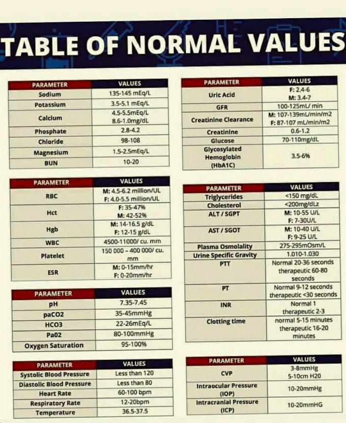
Differential count
|
Normal range
|
|
Neutrophils
|
40-72%
|
(1800-7800/
|
Eosinophis
|
1-6%
|
(50-450/µl)
|
Basophiles
|
0-0.5%
|
(0-200µl)
|
Lymphocytes
|
20-45%
|
(1000-4500/µl)
|
Monocytes
|
1-8%
|
(100-800/µl)
|


If you have any queries related medical laboratory science & you are looking for any topic which you have have not found here.. you can comment below... and feedback us if you like over work & Theory
.
Thanks for coming here..