MORPHOLOGY AND STRUCTURE OF BACTERIA
The classification of bacteria in routine use is
'artificial' and is based on some recognizable features and characteristics
which have been arbitrarily selected. These features include:
- Morphology
- Staining reaction
- Cultural requirements
- Biochemical reactions
- Antigenic structure
- DNA base composition i.e. guanine: Cytosine ratio
 |
| Morphology And Structure Of Bacteria |
Morphology and Anatomy of Bacteria
what is Bacteria
Bacteria are microscopic unicellular organisms. The unit of
measurement of bacteria is the micrometer (um) and it is 1/1000 of a millimeter
(.001 um). Morphological classification of bacteria is based on the following
types of shapes of cells:
1. The spheroid or
ovoid or coccus
2. Bacillus
3. Vibrio
4. Spirillum
5. Spirochaete
1. The spheroid or
ovoid or coccus These groups of bacteria are popularly called cocci (plural
of coccus). They measure about 0.5-1 um in diameter. Sometimes these cells are
flattened or distorted to give a change in shape.
- The bacteria multiply by binary fission. During multiplication, the daughter cell is attached to the parent cell but it gets detached before fission occurs again. The pairs of cocci seen are referred to as diplococci.
- If the fission continues while they remain attached, chains of cocci are formed and are referred to as streptococci. If the division is not in one plain and clusters of cocci are formed randomly, they are known as staphylococci.
- Tetrads (or tetracocci) are formed when the cocci remain in pairs for two consecutive divisions and form regular aggregates of four cocci.
- When the terms Staphylococcus and Streptococcus are used as generic names, the first letter is a capital letter.
2. Bacillus This
is a 'rod' shaped cell measuring about 1-10 um in length and 0.3-1.0 um in
width. The bacilli (plural) or rods may remain attached after division and they
are called streptobacilli; or they may be arranged at varying angles to one
another due to incomplete separation after cell division, thus giving the
appearance of Chinese lettering. This is characteristic of the genus
Corynebacterium.
The morphology of some of these bacilli may be affected as a result of the formation of spores during unfavorable conditions. Some species contain these spores in the center with or without bulging and others may have them at terminal ends or towards one end.
3. Vibrio The
vibrio is a short, comma-shaped rigid bacillus. It measures about 4 x
5um.
4. Spirillum The
spirillum is a rigid spiral organism and it is variable in size, measuring
approximately 4 x 0.2 um. It has a corkscrew (helical) shape.
5. Spirochaete
This is a flexible spiral-shaped organism. It possesses an axial fiber around
which the body is twisted in a helical fashion. It measures about 10–20 um in
length and 0.2-0.4um in thickness.
Figure 2.1 shows the various bacterial shapes and
arrangements.
 |
| Structure Of Bacterial Morphology |
 |
Anatomy of Bacteria
|
Morphology And Structure Of Bacteria
1. Flagella These
are slender filaments that originate from the cytoplasm. They function as
organs of motility. They are therefore seen only in organisms that are motile.
They are proteins and have characteristic patterns of arrangement on the
bacterial cell (Fig. 2.3). They are too thin to be seen in ordinary stained
preparations but become visible only in silver impregnation preparation or in
electron microscopy.
2. Pili Extruding
from the cytoplasmic membrane is shorter and finer filaments than the
flagella. These are the pili. Some of them are referred to as sex pili because
of their role during conjugation when genes are transferred from one cell to
another. Majority of them are referred to as common pili or fimbriae. These are
thought to be the organs of adhesion that help bacteria to attach to the host
cells.
3. Capsule This
is a layer of loose slimy material that surrounds some bacterial cells. The
capsules are composed mainly of polymers of polysaccharides or peptides. They
resist phagocytosis and so their presence on a bacterium is associated with
virulence. They are identified by negative staining due to their low affinity
for simple stains.
 |
| Distribution of flagella |
4. Cell Wall The
cell wall is a prominent distinguishing feature of the bacterial cell with a
rigidity and strength that protects the cellular contents. Chemically, the cell
wall consists of peptidoglycan, a mucopeptide that is composed of alternating
strands of N-acetyl-glucosamine and N-acetylmuramic acid residues, cross-linked
with peptide subunits.
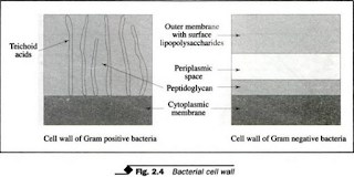
- The peptidoglycan gives rigidity and shape to the organism. Structurally, the cell wall of a Gram-positive bacterium differs from that of a Gram-negative bacterium (Fig. 2.4). The Gram-positive cell wall is much more sturdy than the Gram-negative one due to its high content of peptidoglycan.
- It has neither an outer membrane that contains specific proteins nor a periplasmic space, both of which are present in the Gram-negative cell. Teichoic acids are also part of the cell wall of Gram-positive bacteria.
- The Gram-negative cell wall has a thin peptidoglycan layer with no teichoic acids. It has an outer membrane that consists of lipopolysaccharides. The lipopolysaccharide is toxic to humans. It is called endotoxin.
 |
| Bacterial cell wall |
5. Cytoplasmic
membrane This is a double-layered structure within the cell wall. It is
composed of lipid and protein and acts as a semipermeable membrane through
which nutrients pass into and out of the cell.
INTERNAL STRUCTURES
1. Mesosomes
These are convoluted invaginations within the cytoplasmic membrane. They play
an important role during cell division and in the secretion of certain
enzymes.
2. Nuclear material
Within the cytoplasmic membrane, the cell itself has the nucleus' which has no
nuclear membrane and lacks a definite shape. It is a single circular strand of
deoxyribonucleic acid (DNA) which acts as a 'nucleus (chromosome).
3. Ribosomes
Ribosomes are located throughout the cytoplasm and are the sites of protein
synthesis. They are important for conveying the genetic code of the nucleus
into instructions in the manufacture of cellular components. They are composed
of ribonucleic acid (RNA) and proteins.
4. Cytoplasmic
inclusions These are seen in some bacteria and appear to be sources of
reserved food for energy. For example, volutin (polymetaphosphate) granules
associated with Corynebacteria.
5. Spores These
are dense structures produced by the bacteria, e.g., the Bacillus and
Clostridium groups, that enable them to survive adverse environmental
conditions. They develop within and at the expense of the vegetative cell.
- The spore comprises the chromosomal material surrounded by several layers of walls. The location and shape of the spore in the cell may be of diagnostic assistance, e.g, the spores of Clostridium tetani are terminal, and the diameter is greater than that of the parent cell so that they are characteristically of drum stick appearance.
- The positions of spores are described as terminal, subterminal or central. Spores are resistant to heat, stains, desiccation, and disinfectants. Each spore germinates to produce a vegetative cell during favorable growth conditions.
 |
| Morphology And Structure Of Bacteria |


If you have any queries related medical laboratory science & you are looking for any topic which you have have not found here.. you can comment below... and feedback us if you like over work & Theory
.
Thanks for coming here..