MORPHOLOGICAL VARIATIONS/ ABNROMALITIES IN ERYTHROCYTES
MORPHOLOGICAL VARIATIONS/
ABNROMALITIES IN ERYTHROCYTES-Alterations in the morphology of
erythrocytes are associated with many diseases. The most significant of these
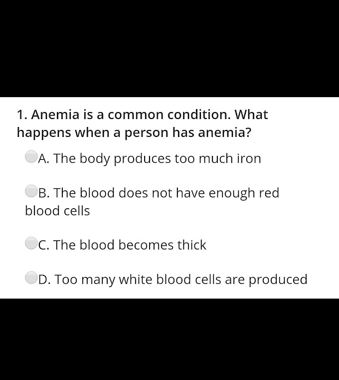
conditions is anaemia, in which the oxygen carrying capacity of blood is
decreased. The causes of anaemia are varied, and most of them are expressed as
changes in RBC morphology. Therefore, morphological examination of red cells is
very helpful in evaluating and determining the cause of anaemia.
Normal red blood cells stained with a Romanowsky stain are
nearly uniform in size, shape and colour. Each cell appears as a pink disk,
about 7 microns in diameter, with a rim of haemoglobin and a clear central area
called central pallor. The central pallor generally occupies less than
one-third of the cell. The red cells having normal size and normal colour are
said to be normocytic and normochormic. Blood disorders may also be indicated
by changes in the shape of red cells or by the presence of inclusions. Figure
4.4 shows some red cell abnormalities and inclusions.
During the examination of the
peripheral blood smear, the following characteristics in the red cell
morphology should be observed and reported:
1. Colour (Haemoglobin content)
2. Size
3. Shape
4. Inclusions
5. Distribution pattern
6. Nucleated red cells
7. Artefacts.
1. Variation in Colour:-A red
cell showing a normal staining reaction is described as normochromic and
represents a haemoglobin content within the normal range.
Hypochromic cells stain very
pale and show an increased area of central pallor. Hypochromasia is a result of
reduced haemoglobin content, most commonly observed in iron-deficiency anaemia.
Hyperchromic cells are not seen
very commonly. These cells stain deeply appearing oversaturated with
haemoglobin. Such cells are observed in spherocytosis.
Polychromatic cells show a
mixed staining reaction and appear blue-orange in colour. They contain residual
ribonucleic acid (RNA) which is basophilic, in addition to haemoglobin. These
are slightly premature cells, just released in circulation from the bone
marrow.
A normal blood smear may show 1-2 % polychromatic cells.
They are called reticulocytes. They are slightly larger in size than normal red
cells. When stained with a supra-vital stain such as brilliant cresyl blue,
these cells show a reticulum of RNA. An increase in the number of these cells
in the peripheral blood indicates increased red cell production by the bone
marrow and may be seen in conditions such as haemolytic anaemia.
2. Variation in Size:-
Variation in size, called anisocytosis, is a common and important erythrocyte
morphological feature. The term describes any large variation in size of a
normal erythrocyte. A red cell within the normal range of diameter is called a normocyte.
Macrocytosis:_ It is a condition in
wnich red blood cells have a diameter greater than 7.8 microns and MCV usually
greater than 100 cu.microns (femtolitres, fL). Macrocytic cells are
characteristically seen in megaloblastic anaemia due to Vit B12 or folic acid
deficiencies, or in haemolytic anaemia.
Microcytosi:-s It is a condition in which
red blood cells have a diameter less than 6.5 microns and MCV less than 80 cu
microns(fL). Microcytic cells frequently have less haemoglobin than normal
cells and are seen in iron deficiency anaemia, spherocytic anaemia, lead
poisoning and thalassaemia.
3. Variation in Shape:-A normal
red cell is a circular, biconcave disc. Any alteration in this shape should be
observed.
Poikilocytosis:- The term describes a condition in which there
are major variations in the shape of the erythrocyte. The most common of these
variations is a tear drop shape. They are found in various anaemias and
haemolytic states.
Spherocyte:- It is a red cell
whose average thickness has increased, usually with a reduced diameter. The
spherical shape results when a normal cell volume is enclosed within a reduced
surface area. When seen in a blood smear, it is a small, deeply staining cell
with no red cell central pallor. A spherocyte has a 14 day life span as
compared to 120 days of a normal blood cell. It commonly shows in creased
osmotic fragility. Spherocytes are found in congenital and acquired haemolytic
anaemias.
Elliptocytes :- These are red
blood cells which are oval or egg-shaped, sometimes almost cylindrical. They do
not show the central pallor. Large elliptocytes may be seen in megaloblastic
anaemias. Elliptocytosis may be acquired or inherited
.
Target cells:- These cells have
a central area of haemoglobin within the area of central pallor, making them
look like targets. This is a result of having excessive cell membrane, compared
to the amount of haemoglobin. They occur in various types of anaemias, liver
disease and after splenectomy.
Sickle cells:-These elongated
erythrocytes with crescentic shapes and occasionally U, S, or L shapes are
formed after exposure to reduced oxygen tension. This variation occurs if the
red blood cell contains the abnormal haemoglobin S. This is a hereditary
disorder, which may appear in two forms: the sickle cell trait or sickle cell
disease.
Burr cells:- These erythrocytes
have a focal crenation producing irregular contractions resulting in long
spine-like processes, irregularly distributed over the cell surface. They have
been observed in renalinsufficiency and thrombocytopenia.
Schistocytes:- These are
fragmented portions of red cells which appear in various shapes, e.g., helmet
cells. Their presence indicates a very serious pathological condition e.g.
mechanical fracture of cells during circulation in conditions such as a
defective heart valve and glomerular filtration. They may also form due to
toxic or metabolic injury as in malignancies. They are frequently seen in blood
smears of severely burnt patients, and in some haemolytic anaemias.
Acanthocytes:- These cells show
irregular margins with pointed projections. The cause of this variation is not
clearly understood, but may be due to an abnormality in the phospholipid
content of the red cell membrane.
Table 4.3 shows causes of variations in shape, size and
colour of red cells.
------------------------------------------------------------------------------------
4. Abnormal Inclusions in Red Blood
Cells:- A normal red cell does not contain any inclusion. Various types
of inclusions may be seen in RBCS which may indicate disorders or disease
condions. Basophilic stippling The red cells may show fine or coarse dark blue
granules dispersed throughout the cell. This is known as basophilic stippling
and results from the precipitation of ribosomal RNA.
The stippling may occur due to abnormal red cell formation
in the bone marrow and is seen in heavy metal poisoning (lead, mercury or
bismuth), thalassaemia and megaloblastic anaemia. Howell-Jolly bodies- These
are round, dense purple granules less than 1 micron in size, appearing
eccentrically located in some red cells. If present, not more than two
Howell-Jolly bodies are seen in a single red cell. They are the remnants of the
nuclear material and indicate incomplete expulsion of the nucleus from the red
cell through the spleen. Therefore, the Howell-Jolly bodies may appear in the
peripheral blood after splenectomy, and also in megaloblastic anaemia and some
haemolytic anaemias.
Heinz bodies:- Heinz bodies are
oxidised denatured haemoglobin. They can be demonstrated only by supravital
staining with brilliant cresyl blue. Heinz bodies my appear in the peripheral
blood smear after removal of the spleen, or in haemolytic anaemias such as that
due to G6PD deficiency.
Siderocytes:- The red cells
containing small, dense, blue-purple granules are called siderocytes. They
resemble Howell-Jolly bodies in appearance, but they are granules of free iron,
uncombined with haemoglobin, instead of fragments of DNA. The iron-granules
inside siderocytes are known as Pappenheimer bodies. A special stain for iron,
such as Prussian blue, can distinguish these granules from Howell-Jolly bodies.
They are seen in peripheral blood after the removal of spleen.
Cabot's rings:- These are rare
inclusions appear ing as threadlike strands in the form of a ring or figure of
8, reddish-violet in colour. Their origin is not clearly understood, but may be
the result of anabnormality in mitosis. They appear in cases of megaloblastic
anaemia or lead poisoning. They can be differentiated from the malarial ring
forms by their large size and absence of red chromatin.
 Parasites:- Various stages of
the malarial parasites may be seen in the red cells of patients suffering from
malaria. Their appearance depends on the developmental stage (e.g. ring form,
amoeboid trophozoite, schizont, gametocyte) and the infecting species of the genus
Plasmodium, e.g., Plasmodium vivax, Plasmodium falciparum. Other parasites such
as trypanosomes and microfilariae may also be seen in a peripheral blood smear.
Blood parasites are discussed in greater details in Parasitology in
Microbiology Section.
5. Alterations in the Distribution
Pattern of Red Cells
Rouleaux Formation:- Sometimes
red cells stick together in row looking like stacks of coins. This is called
rouleaux formation. They may appear in the thick portion of the smear of
healthy individuals. However, their presence in the thinner 'examination area
of the smear indicates increased levels of plasma globulin or fibrinogene.g. in
multiple myeloma
Agglutination:- Agglutination
is irregular clumping. If agglutination is observed in blood samples stored in
the refrigerator, it may be due to the presence of cold agglutinins, which are
autoantibodies indicating an auto-immune haemolytic state or anaemia.
6. Nucleated red Cells in the
Peripheral(Blood (Erythroblastic Reaction):-The peripheral blood of a
healthy individual does not show the presence of nucleated red cells. Their
appearance in the peripheral blood indicates intense stimulation of the bone
marrow releasing red cell precursors in the peripheral blood. This occurs in
acute blood loss, megaloblastic and haemolytic anaemias and malignancies
Normoblasts (erythroblasts) in various stages of development may be seen
depending on the severity of the stimulus.
7. Artefacts in Red Cells:-
Platelets on top of a red cell, punched-out red cell or stain deposits may
appear as variations in red cell morphology . they should not they should be
confused with red cell abnormalities.


If you have any queries related medical laboratory science & you are looking for any topic which you have have not found here.. you can comment below... and feedback us if you like over work & Theory
.
Thanks for coming here..