MICROSCOPIC EXAMINATION OF URINE
 The most
common laboratory procedure utilised for the detection of renal or urinary
tract disease is the microscopic examination of urine. This can be done in
various ways, but is generally carried out by taking a standard amount of
urine, centrifuging the specimen for a fixed period of time, removing the
supernatant fluid, resuspending the sediment in a standard volume (usually 1
ml), and examining the sediment under the microscope.
For
semiquantitative or standardised analysis of the urinary sediment, a constant
volume of urine must be centrifuged and a constant volume of supernatant
removed. For example, 12 ml of urine should becentrifuged and finally
resuspended in 1ml. Examination of a urinary sediment enables assessment of the
number of cells (white cells, red cells, epithelial cells, and tumour
cells),casts, crystals and parasites in urine to be done. Each of these
constituents can be a valuable indicator of pathology.
There are various ways in which a urinary sediment can be examined. With a proper level of professional expertise, bright-field microscopy of unstained urine can identify all clinically important and relevant structures in the sediment. It is necessary to adjust the amount of light entering the objective by carefully positioning the condenser and iris diaphragm. Other methods of examination include phase-contrast, interference or polarised microscopy or use of staining methods such amonochrome staining, gram staining or supra-vital staining.
Constituents of Urinary Sediment
The constituents of urinary sediment can be
broadly divided into two groups:
1.
Organised sediment This represents the biological part and includes various
types of cells, casts, bacteria, parasites and spermatozoa. Cells in the urine
are either from the kidney or urinary tract, or from circulating blood.
Organisms and tumour cells do not originally belong to the urinary system
7.URINARY CALCULI:
Method for Microscopic Examination of
Urine
For
standardisation of reporting the urinary sediment is concentrated 12:1, that is
12 volumes of urine is centrifuged, supernatant removed and the sediment resuspended
in 1ml of urine before examination. Any fraction of this ratio may be used
(e.g., 6 ml to 0.5 ml, 3 ml to 0.25 ml)
Method
1. Pour 12
volumes (ml) of well-mixed urine in a centrifuge tube and centrifuge at 450 g
(approx. 1500-2000 rpm) for 5 minutes.
2. Decant 11
volumes (ml) of urine without disturbing the sediment, leaving 1 volume (ml) in
the tube.
3. Gently
resuspend the sediment. Deliver one drop on a slide. Cover with a coverslip.
4. Examine
the slide using low power (10 X) objective. Reduce the light intensity to
minimum and scan several fields for casts. If present, report the number of
cast per low power field. Identify the cast type by using high power (40 X)
objective.
5. Using
high power objective with slightly increased light intensity, identify and
count red cells, leukocytes and renal epithelial cells. Examine at least 10
fields. Report each cell type as cells/hpf.
6. Report on
(i)
Bacteria, yeast, parasites, fat globules if present as few, moderate, many or
abundant. Spermatozoa should be reported if present in large numbers
(ii)
Crystals: Identify and quantitate as few,moderate or many. Presence of abnormal
crystals should be confirmed chemically before reporting.
(iii)
Squamous and transitional epithelial cells if present in large numbers.
(iv) Large
amounts of mucus.
(V) Any
other abnormal finding.
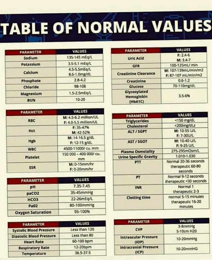
Note - 0-5 leukocytes per hpf, 0-2 red
cells per hpf, and 0-2 hyaline casts per Ipf are considered to be within normal
limits.


If you have any queries related medical laboratory science & you are looking for any topic which you have have not found here.. you can comment below... and feedback us if you like over work & Theory
.
Thanks for coming here..