ERYTHROCYTE SEDIMENTATION RATE (ESR):-
ESR, though not a part of Complete Blood Count, is usually
requested with it. The basic principle of the ESR is based on the fact that
blood is essentially a suspension of formed elements such as red and white
corpuscles in plasma.
Therefore, when whole blood is mixed with an anticoagulant and placed in a perpendicular tube, the red blood cells sink to the bottom because they are heavier than the plasma in which they are suspended. The speed at which the red blood cells in normal blood settle is relatively slow.
However, in many diseases, e.g.. inflammatory or degenerative cell diseases, changes occur in Physico-chemical properties of the plasma. These changes include alterations in the positive charges of the plasma colloids, increase in plasma fibrinogen, variation in the concentration of plasma protein fractions and in the ratio of the various plasma protein fractions to one another.
Changes may also occur in the erythrocyte surface.
Change in the surface electric charge of the red blood cells causes the erythrocytes to aggregate, clump, or to form rouleaux. The large clumps of cells thus formed fall at a faster rate. The changes in the proportion of the soluble constituents of plasma such as increased fibrinogen or globulin also result in an increased rate of erythrocyte fall.
Change in the surface electric charge of the red blood cells causes the erythrocytes to aggregate, clump, or to form rouleaux. The large clumps of cells thus formed fall at a faster rate. The changes in the proportion of the soluble constituents of plasma such as increased fibrinogen or globulin also result in an increased rate of erythrocyte fall.
Sequentially, there are 3 stages in erythrocyte sedimentation.
1. The initial period
of a few minutes, during which rouleaux formation occurs.
2. A period of approximately 30 to 120 minutes, depending on the tube length, during which sedimentation occurs at a fairly constant rate.
2. A period of approximately 30 to 120 minutes, depending on the tube length, during which sedimentation occurs at a fairly constant rate.
3. A period of a slower rate of fall during which packing of
the sedimented red cell column occurs. For these reasons, the sedimentation rate
test must be set up within 2 to 6 hours of blood collection.
There are two
main methods of performing the ESR, viz., Wintrobe's and Westergren's methods.
1.Wintrobe's methods for ESR
2. Westergren Method for ESR
3. Micro ESR Method for ESR
Westergren Method for ESR (Erythrocyte Sedimentation Rate)
 |
Westergren Method for ESR (Erythrocyte Sedimentation Rate)
|
1.
Westergren ESR tube This tube looks similar to a one ml pipette. It is 300 mm
long with an internal diameter of 2.5 mm. It is graduated from the base over a
200 mm scale in mm divisions. (Fig 3.10)
2. Sodium citrate 3.8 % (W/V)
Specimen need in Westergren Method
Add 0.5 ml of 3.8 % sodium citrate in a small test tube. Collect 2 ml of venous blood after overnight fasting and mix with the sodium citrate. |
| western,s ESR pipette with stand |
Westergren Method Procedure
- Aspirate the blood into the Westergren tube to the top mark (O).
- The tube is then stood vertically in a stop rack with spring clips that hold the lower end of the pipette tightly against a rubber mat in a vertical position.
- Leave for one hour. Note the readings as for Wintrobe's method.
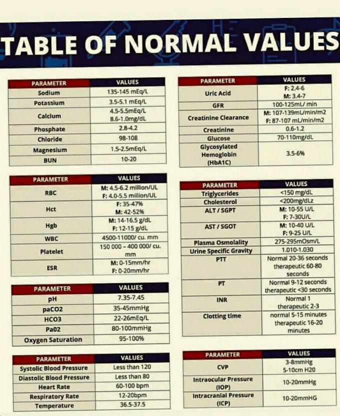
Normal Range At the end of one hour:-
- Normal values
- Males 3-5 mm 1st hour
- Females 4-7 mm 1st hour
Advantages and Disadvantages of Westergren method
Advantages |
Disadvantages |
i. It is a more sensitive method. |
i. It requires more amount of blood.
|
ii. It is easy to fill and clean the Westergren’s
pipette.
|
ii. Dilution of blood in anticoagulants effects ESR.
|
iii. Filling of blood by mouth pipetting should be strictly
discouraged. |
 |
| Westergren Method for ESR |
Wintrobe’s Method for ESR (Erythrocyte Sedimentation Rate).
 |
Wintrobe’s Method for ESR (Erythrocyte Sedimentation Rate)
|
The Wintrobe tube is a glass tube closed at one end. The tube is 110 mm long and has an internal bore diameter of 2.5 mm. The the tube is graduated on both sides from 0 to 10 on one side and 10 to 0 cm on the other
Specimen for Wintrobe’s
5 ml of venous blood after an overnight fast is withdrawn and immediately mixed in a tube containing 6 mg of dried ammonium oxalate and 4 mg of dried potassium oxalate.Anticoagulants Either of the following 2 anticoagulants can be used
i. Ethylenediaminetetraacetic acid (EDTA) solid crystals 1-2 mg/ml.ii. Double oxalate (solid) 2-3 mg/ml(ammonium oxalate and sodium or potassium oxalate in the ratio of 3:2; the former causes swelling and the latter causes shrinkage of RBCs and hence RBC shape is retained).
 |
| WINTROBE'S TUBE AND PASTEUR PIPETTE |
Requirements-
Wintrobe hematocrit tube This tube is 11 cms in length with an internal diameter of 2.5 mm. It is graduated from the bottom in mm divisions over a 10 cms scale. When filled, the tube contains 0.7 ml of blood.the procedure of Wintrobe’s Method
- The the patient is called in the morning fasting.
- Draw 1 ml of blood into the anticoagulant.
- Fill the Wintrobe tube up to mark 0 with anticoagulated blood with the help of a Pasteur pipette having a long stem (15 cm) so as to fill the tube free of air bubbles
- Place the tube vertically in a stand and note the ESR after one hour.
OR
- Using a Pasteur pipette with a long stem.
- fill the hematocrit tube to the 10 mark with well-mixed blood.
- Forming of air bubbles in the tube should be avoided.
- Fix the tube in the stand, ensuring that the tube is vertical.
- Take readings preferably at 15-minute intervals for one hour in order to get an indication of the sedimentation rate.
- Note the level of red cells from 0 at the top.
The normal range of ESR by Wintrobe’s Method
- Males 0-7 mm 1st hour
- Females 0-15 mm 1st hour
Advantages and Disadvantages of Wintrobe’s method
Advantages |
Disadvantage |
i. It
is a simple method and requires a small amount of blood.
|
i. Because of the short column and choice of
anticoagulant, it is not as sensitive index of diseases.
|
ii. There is no dilution with an anticoagulant.
|
ii. Addition of more anticoagulant can lower ESR. |
iii.
Packed cell volume (PCV) can also be done by the same tube.
|
iii. ESR of more than 100 mm can not be
measured.
|
iv. Filling of a tube with Pasteur pipette
eliminates the chance of any infection due to the handling of blood.
|
Micro ESR Method for ESR (Erythrocyte Sedimentation Rate)
 |
| Micro ESR Method for ESR (Erythrocyte Sedimentation Rate) |
This method is used in pediatric patients or in patients where
venepuncture is not possible. In this method, a capillary 160 mm long with an
internal bore diameter of 1 mm is used in the micro ESR method.
- The capillary is graduated 1 mm apart for 50 mm, with two red lines on it. Alternatively, non-graduated heparinized capillary may be used and the reading is taken by measurement of length of column
Specimen for Micro ESR Method
- Anticoagulant Mixture of sodium citrate and EDTA is used.
Procedure for Micro ESR Method
 |
| Micro ESR Method for ESR |
- Fill the micro sedimentation pipette up to the first red mark with an anticoagulant.
- Fill the pipette with free-flowing capillary blood up to second red mark.
- Invert it several times and allow it to stand for one hour in the sedimentation rack.
- Take the reading and results are given as that for Westergren Method.
Normal Range At the end of one hour:-
- Normal values
- Males 3-5 mm 1st hour
- Females 4-7 mm 1st hour
Clinical Significance of ESR
ESR is a non-specific method of evaluating diseases. It is
seldom used for diagnostic purpose but its use is limited to monitoring the
prognosis of the disease process.
Diagnostic
Uses of ESR
i. Rheumatoid arthritis
ii. Chronic infections
iii. Collagen diseases
iv. Multiple myeloma
v. Macroglobulinaemia
Diagnostic Uses of ESR
i. Rheumatoid arthritis
ii. Chronic infections
iii. Collagen diseases
iv. Multiple myeloma
v. Macroglobulinaemia
Monitoring Prognosis of Diseases To see the response to treatment in: |
i. Tuberculosis
|
ii. Temporal arteritis
|
iii. Polymyalgia rheumatica
|
iv. In patients of Hodgkin’s disease, ESR of <
10 mm
1st hour indicates good prognosis while ESR of
> 60 mm 1st hour indicates poor prognosis.
|
Diseases causing Diseases ESR |
Tuberculosis
|
SABE
|
Acute
myocardial
|
infarction
|
Liver
disease
|
Multiple
myeloma
|
Pregnancy
|
Rheumatoid
arthritis
|
Causing raised ESR low |
i. Polycythaemia
|
ii. Spherocytosis
|
iii. Sickle cell anemia
|
iv. Congestive heart failure
|
v. Newborn infant
|
vi. Hypofibrinogenaemia
|
High ESR indicates
- Infection
- Rheumatoid arthritis
- Rheumatic fever
- Vascular disease
- Inflammatory bowel disease
- Heart disease
- Kidney disease
- Certain cancers
Main methods of performing the ESR
High ESR indicates
- Infection
- Rheumatoid arthritis
- Rheumatic fever
- Vascular disease
- Inflammatory bowel disease
- Heart disease
- Kidney disease
- Certain cancers


If you have any queries related medical laboratory science & you are looking for any topic which you have have not found here.. you can comment below... and feedback us if you like over work & Theory
.
Thanks for coming here..