CSF Analysis and Other Body Fluids
Formation of Cerebrospinal Fluid (CSF)
Functions of CSF
Collecting CSF
Possible complications of lumbar puncture,
Indications for CSF examination
Composition of CSF
ROUTINE LABORATORY EXAMINATION OF CSF
CEREBROSPINAL
FLUID (CSF)--The investigation of clinical disorder of the central
nervous system (CNS) is partly based on the examination of cerebrospinal fluid
(CSF). The central nervous system consists of the brain, spinal cord and
peripheral nerves.
The brain is encased within the cranial cavity and weighs
about one-fifth of the body weight. Structurally it is partitioned into the
cerebrum (greater brain), the brain stem (midbrain, pons varolii and medulla
oblongata) and the cerebellum (lesser brain)
 |
| CEREBROSPINAL FLUID (CSF) |
1. An outer protective
membrane called the duraa mater;
3. An inner membrane called the pia mater (Fig. 10.1b).
The arachnoid mater and the pia mater are separated from
each other by the subarachnoid space which is filled with the cerebrospinal
fluid. Thare are subdural space Between the duraa mater and the arachnoid
mater.
Formation of Cerebrospinal Fluid (CSF)-
 |
Formation of
Cerebrospinal Fluid (CSF)
|
The fourth ventricle lies between the pons and cerebellum of
the brain. The CSF then flows from the fourth ventricle down the small central
canal of the spinal cord and through openings (foramina) into the subarachnoid
space to completely surround the brain and the spinal cord. Small blood vessels
in the arachnoid mater constantly reabsorb CSF in circulation so that the
production is balanced by an equal reabsorption of fluid.
Functions of CSF (Cerebrospinal Fluid)
1. Cerebrospinal Fluid provide a "fluid cushion"
to support and protect the brain and the spinal cord.
2. It acts as a shock absorber for the brain and the spinal
cord.
3. It carries nutrients to the brain and spinal cord and
removes waste products.
4. It helps to maintain a constant pressureinside the head
and around the spinal cord.
5. It keeps the brain and spinal cord moist.
Collecting
CSF (Cerebrospinal Fluid)
A specimen of
CSF is collected by a lumbar puncture, using a long needle with a stylette
inside. The needle is introduced between the third and fourth lumbar vertebrae
into the spinal sub-arachnoid space with the patients back well flexed for good
separation of the vertebrae. It is a very critical operation and damage to the
spinal cord can easily occur.
The cord stops at the
level of the first lumbar vertebra and cannot be damaged by the needle entering
the subarachnoid space a few centimetres lower.
The CSF is usually collected into 2 or 3 sterile containers,
numbered according to the order of collection, each containing about 1-3ml of
CSF.
Possible complications of lumbar puncture
1. Production of cerebellar pressure cone in patients with
increased intracranial pre ssure.
2. In the case of spinal cord tumour, paresis may progress
to paralysis as a result of lumbar puncture.
3. During the whole process of lumbar puncture, infection
can inadvertently be introduced by
(a) the use of unsterile equipment; (b) by passing the
needle through a septic spot in the lumbar region,(c) by not observing aseptic
precautions.
4. After-puncture headache as a result of leakage of CSF.
This condition can be minimised by using a small bore needle.
Indications for CSF examination
1. Diagnosis and detection of suspected meningitis,
subarachnoid haemorrhage, encephalitis, central nervous system syphilis, spinal
cord tumour or multiple sclerosis.
2. Differential diagnosis of cerebral infarction from
intracerebral haemorrhage (about 80 % of the latter show xanthochromia or
bloody appearance of CSF).
Composition of CSF(Cerebrospinal Fluid):-
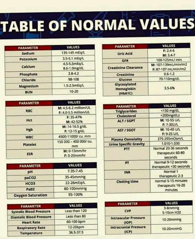
The CSF is mainly composed of water, dissolved oxygen and solids. The average volume is between 100 ml and 150 ml which is produced at the rate of 430 ml/day. It is similar to plasma in composition, but the concentrations of sodium, chloride, magnesium and glutamine are greater in CSF than in plasma; while concentrations of glucose, potassium, calcium, cholesterol, uric acid and iron are higher in plasma. Table 10.1 shows the composition of normal CSF.Compostion of normal Cerebrospinal Fluid (CSF) |
|
Volume
|
100-150ml
|
Total
protirn
|
0.15-0.45g/dl
|
Glucose
|
3-4.5 mmol/l (50-80mg%)
|
chloride
|
118-134 mmol/l
|
Sodium
|
144-154 mmol/l
|
PH
|
7.3-7.4
|
CELLS
|
0-5 Lymphocytes/cu.mm
|
ROUTINE
LABORATORY EXAMINATION OF CSF(Cerebrospinal Fluid)


If you have any queries related medical laboratory science & you are looking for any topic which you have have not found here.. you can comment below... and feedback us if you like over work & Theory
.
Thanks for coming here..