

ROUTINE LABORATORY EXAMINATION OF CSF
-Estimation of total proteins in CSF. by Turbidimetric,

Chemical
Examination of CSF
Trichloroacetic, Micro method,
-Estimation of Globulin in CSF by Pandy's test
Nonne-Apelt's- method, Radial immunodiffusion test,
-Clinical Significance CSF proteins,
-Desease condition that effect the CSF glucose levels,
-Bacteriological Examination of CSF
-Bacteriological examination of synovial fluid,
-Mucin Clot test evaluate Viscosity of Synovial Fluid

Chemical Examination of CSF
Chemical Examination of CSF
Chemical Examination of CSF, Estimation of total proteins in
CSF. by Turbidimetric methods ,
Trichloroacetic Acid method ,Micromethod
Estimation of Globulin in CSF by Pandy's test, Nonne-Apelt's- method , Radial
immunodiffusion test
Clinical Significance CSF proteins
Desease condition that effect the csf glucose levels
Bacteriological
Examination of CSF
SYNOVIAL FLUID
Becterialogical examination of synovial
fluid,
Viscosity of Synovial Fluid
Mucin Clot test evaluate the Viscosity is
in the laboratory of Synovial Fluid
Proteins-
Turbidimetric methods are commonly employed for the estimation of total
proteins in CSF.
Turbidimetric methods
Principle- Proteins in CSF are precipitated by either dilute trichloroacetic acid or
sulphosalicylic acid in sodium sulphate solution. The turbidity of the
resultant uniform suspension is measured in a colorimeter against a known
standard.
Trichloroacetic Acid method
Reagents
1. Trichloroacetic acid 3% aqueous solution.
2. Stock standard protein solution 5.0 g/L in 0.90% NaCI:
Distribute in small volumes and store at -18°C. The standard can be prepared
from diluted pooled serum, or making suitable dilutions of commercial control
serum or from bovine albumin.
3. Working standard protein solution 0.5 g/L (50 mg %):
Dilute the stock standard 1 in 10 with 0.9% NaCl. Fresh stock is to be prepared
each week and stored in 1.5ml volumes at about -18°C. Thaw a sample for use
each day.
Method
1. To 4.0 ml of trichloracetic acid, add 1.0 ml of CSF drop
by drop with constant mixing.
2. In the same manner, add 1.0 ml of standard to 4.0 ml of
trichloroacetic acid.
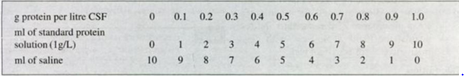
Preparation of calibration curve --Dilute the stock standard
protein (5.0 g/L) 1 in 5 with 0.9% NaCl (saline) and prepare a series of tubes
as follows:
3. Add 1.0 ml distilled water to 4.0ml trichloroacetic acid.
This is the blank.
4. Stand at room temperature for 10 minutes and remix the
turbid solutions.
5. Read the absorbances of the standard and test against the
blank at 450 nm or by using a blue filter.
Calculations
T= Absorbance of Test;
S = Absorbance of
standard
T/S x0.5 = g CSF protein per litre
To convert to mg/100 ml (mg %), multiply by 100.
Note
1. Use appropriate dilutions if the value of the test
exceeds the upper limit of the method as established by a cali bration curve.
2. A calibration curve will show if a linear relationship
holds for increasing protein concentration and should be prepared for each
photometric in strument.
3. The method is temperature depend Ent
4. The size and shape of the particles affect the
absorbance. The tubes must be shaken to mix constantly for up to 4 times.

Mix well; add 1.0 ml of each standard protein solution to
4.0 ml trichloroacetic acid. Read the absorbance at 450 nm (blue filter). A
straight line calibration curve is obtained if the absorbance is linear.
Method using permanent standard This method is not very accurate but it is such a rapid and simple method that most bacteriology laboratories which are not usually equipped for chemical procedures, find it of adequate accuracy.
Reagents 1. 3% Sulphosalicylic acid.
2. Permanent protein
standard marked in mg100 ml (10-100 mg/100 ml) supplied commercially.
Method
1. Add 1.0 ml of clear CSF to 3.0 ml of sulphosalicylic acid
in a standard test tube.
2. Mix contents and leave for 5 minutes.
3. Compare the tube with the turbidity standards Results
Normal CSF contains protein between 0.15-0.45 g/L ( 15 to 45 mg/100 ml). If the
value of the CSF protein is higher than 100 mg/100 ml, dilute with saline and
adjust the result accordingly.
Micromethod- This method is very useful
when the volume of the CSF is small.
Reagent 3% aqueous trichloroacetic acid.
Method 1.
Set up 3 test tubes as follows:
Blank: 0.2 ml of saline Standard: 0.2 ml of standard protein
Test: 0.2 ml of CSF
2. Add to all tubes 0.8 ml of 3.0% trichloroacetic acid. Mix
by inverting about 5 times. Allow to stand for 10 minutes. Read absorbance of
the standard and test against the blank at 450 nm with glass microcuvettes of
Icm light path.
Calculation

Globulin
Tests for globulin are performed only on blood-free CSF
specimens.
1. Pandy's test
Principle-
In a saturated aqueous solution of phenol, globulin molecule absorbs water and
the phenol is displaced from the solution, causing a fine persistent turbidity
Method:- Add 1 drop of CSF to 0.5 ml of saturated
aqueous solution of phenol (must be very clear) in a small test tube.
Cloudiness/turbidity against a dark background indicates increase in globulin
content. Normal CSF remains clear.
Report as Nil, +, ++, +++ or ++++.
2. Nonne-Apelt's- method
Principle-
Globulin is precipitated by solution of saturated ammonium sulphate. Method
Carefully layer 1.0 ml of clear CSF on to 1.0 ml of saturated ammonium
sulphate. A thin white ring of precipitation appears at the interface of the
two liquids. On mixing, the ring may disappear indicating a 1+ reaction. Heavy
cloudiness indicates a 4+ reaction.
3. Radial immunodiffusion test -
--This is a useful test for the measurement of concentrations of albumin and globulin in CSF. This involves the diffusion of an antigen (IgG or albumin) in the CSF through a semi-solid medium containing an antibody (anti-IgG or anti albumin). thereby forming visible zones of precipitation. The measurement of the zones of precipitation corresponds to the concentration of the antigen. This method also determines IgG/albumin ratio which is of diagnostic importance in multiple sclerosis and other neurological disorders.
Note:Electrophoresis
of CSF protein can also be carried out to evaluate albumin and globulin content
in CSF. The Lange's colloidal gold test, though now obso lete, is a good
substitute for electrophoresis.
Normal range:
Normal CSF contains approximately 60% albumin, 8% immunoglobulins and 32% other
proteins.
IgG/albumin ratio: 17.5
IgG index: <0.7
Clinical Significance
Increase in
CSF proteins
(a) Mild increase can be seen in viral meningitis,
neurosyphilis, subdural haematoma, cerebral thrombosis, brain tumour, multiple
sclerosis.
(b) CSF globulins, especially the immunoglobulin IgG, are
raised in cases of multiple sclerosis, neurosyphilis.
(c) Pronounced increase in CSF proteins is seen in acute
bacterial meningitis, tuberculous meningitis, spinal cord tumour, cerebral
haemorrhage and intracranial tumour.
Glucose CSF glucose is estimated in the same way as for
blood glucose usallay csf glucose less then 40mg/100ml is considered as reduced
level.
Desease condition that effect the
csf glucose levels
|
Viral meningitis
Neurosyphilis
Brain or cord tumor
Cerebral thrombosis
Multiple sclerosis
|
no significance
|
|
Cns
leukemia
Subarachnoid
Hemorrhage
|
Modernate reduction
|
|
Bacterial
meningitis
Tuberculosis
meningitis
Fungal
meningitis
|
Marked reduction
|
It is essential that the glucose assay is carried out as soon as possible after collection in order to prevent the action of glycolytic enzymes in the CSF. This may result in the reduction of glucose level.
Bacteriological Examination of CSF
CSF for bacterial examination must reach the laboratory as
soon as possible after collection in a sterile container. The examination
involves mainly
(1) Grams stain and any other stain requested for,
(2) Isolation of
pathogens,
(3) Serology.
Bacteriological examination of CSF is discussed in the
Microbiology Section of this book.
SYNOVIAL
FLUID:-The synovial fluid (SF) is found in each large joint such as the
knee, ankle, wrist, hip, elbow and shoulder. About Iml of the fluid is normally
present.
To aspirate the fluid, aseptic precautions must be observed.
The fluid is commonly sterile. It is collected into sterile. It is collected in to sterle container and then
sent to the laboratory without deley for the microbiological examnination. For
serological tests a chemically clean tube should be used to prevent clotting.
Becterialogical
examination of synovial fluid:- the procedure is similar to that used
for csf.refer to the microbiology section of this book.
Viscosity of Synovial Fluid
Normal synovial fluid will form a tenacious strand about
4-6cm long if allowed to drop from the end of the needle. A strand of less than
3 cm indicates reduced viscosity. Viscosity is evaluated in the laboratory by
the mucin clot test.
Mucin Clot Test
Principle
This test is based on the polymerisation of hyaluronidase. Method Add synovial
fluid drop by drop to about 20 ml of 5% acetic acid in a beaker.
A normal synovial fluid will form a firm clot with clear
surrounding fluid.
A poor clot is surrounded by a cloudy fluid and may fragment
easily. This indicates an inflammatory condition.
The clots can be graded as good, fair, poor or very poor
depending on the firmness of the clot and the clarity of the surrounding fluid.
If no clot forms, only flakes in a cloudy fluid are seen, it is regarded as a
very poor clot.


If you have any queries related medical laboratory science & you are looking for any topic which you have have not found here.. you can comment below... and feedback us if you like over work & Theory
.
Thanks for coming here..